Antioxidant capacity of EGCG vs. Vitamin E VitaminsC(2)

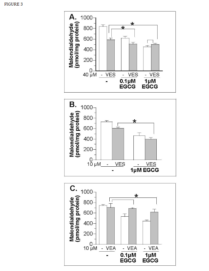
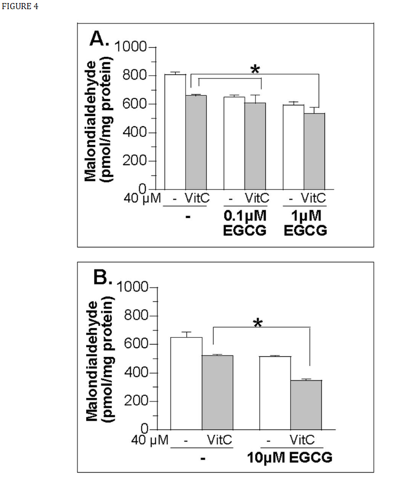
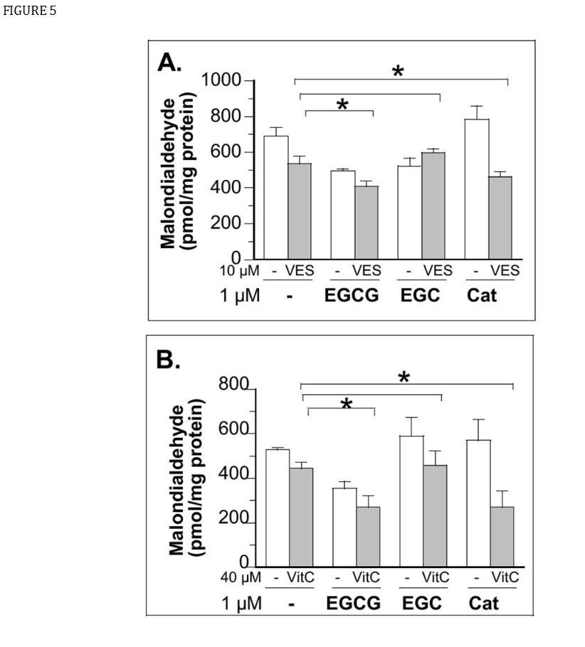
When EGCG was included in the vitamin C preincubation, significantly less MDA production was observed compared to vitamin C treatment alone indicating a higher antioxidant activity (Fig. 4, ,5B5B comparison among gray bars of each panel, left to right with increasing EGCG concentrations in the presence a fixed concentration of vitamin C). Similar to vitamin C experiment, when EGCG was included in cell preincubation medium with vitamin E, significantly less MDA production was observed compared to cells that were treated with vitamin E alone, indicating a higher antioxidant activity (Fig. 3, ,5A5A comparison among gray bars in each panel, left to right with increasing EGCG concentrations in the presence a fixed concentration of vitamin E). Although (±)-catechin does not show lipid antioxidant activity itself, it further decreased the MDA production in cells co-incubated with vitamin C and vitamin E (Fig. 5A and 5B, comparison between the gray bars of vitamin-treated cells in the absence and presence of catechin). Our cell observation is consistent with the prediction and observations made in the cell-free condition. Catechins and phenolic acid were shown to regenerate vitamin E effectively [48-52]. Polyphenols were found to further delay Cu+2-mediated LDL oxidation when added with vitamin C and vitamin E [42]. Feeding catechins has been shown to increase plasma and liver α-tocopherol concentration in rats [52].
In conclusion, our cell studies further support the nutritional value of dietary catechins. In our cell model, EGCG appears to be a more potent lipid antioxidant than vitamin C and E. EGCG also does not have prooxidant activity in physiological achievable concentrations. Although their physiological presence was at a much lower concentration than vitamin C and vitamin E, catechins do contribute additionally to the total lipid antioxidant activity based on our cell studies. Further studies are needed to determine whether our cell culture observation based on non-selective hydroxyl radical attack of lipid can be extrapolated to other types of oxidative stress-induced damages or under in vivo condition.


